
Page 18 - LN-NEURAL CONTROL & COORDINATION
P. 18
bone of the skull. The labyrinth consists of two parts the bony and
membranous labyrinths. The bony labyrinth is a series of channels. Inside the
channels, membranous labyrinth lies, which is surrounded by a fluid called
perilymph.
The membranous labyrinth is filled with a fluid called endolymph. The coiled
portion of the labyrinth is called cochlea.
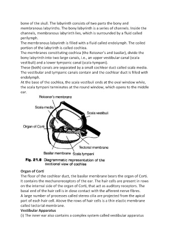
The membranes constituting cochlea (the Reissner’s and basilar), divide the
bony labyrinth into two large canals, i.e., an upper vestibular canal (scala
vestibuli) and a lower tympanic canal (scala tympani).
These (both) canals are separated by a small cochlear duct called scala media.
The vestibular and tympanic canals contain and the cochlear duct is filled with
endolymph.
At the base of the cochlea, the scala vestibuli ends at the oval window while,
the scala tympani terminates at the round window, which opens to the middle
ear.
Organ of Corti
The floor of the cochlear duct, the basilar membrane bears the organ of Corti.
It contains the mechanoreceptors of the ear. The hair cells are present in rows
on the internal side of the organ of Corti, that act as auditory receptors. The
basal end of the hair cell is in close contact with the afferent nerve fibres.
A large number of processes called stereo cilia are projected from the apical
part of each hair cell. Above the rows of hair cells is a thin elastic membrane
called tectorial membrane.
Vestibular Apparatus
(i) The inner ear also contains a complex system called vestibular apparatus