
Page 15 - LN-NEURAL CONTROL & COORDINATION
P. 15
The photoreceptors or visual cells are of two types, i.e., rods (rod cells) and
cones (cone cells). Both of these cells contain light sensitive proteins called the
photopigments.
The twilight (scotopic) vision is the function of the rods. These cells contain a
purplish-red protein called the rhodopsin (visual purple), which contains a
derivative of vitamin-A.
The daylight (photopic) vision and colour vision are functions of cones. There
are three types of cones, which possesses characteristic photopigments that
respond to red, green and blue lights.
The sensation of different colours are produced by various combinations of
these cones and their photopigments. In case of equal stimulation of these
cones, a sensation of white light is produced.
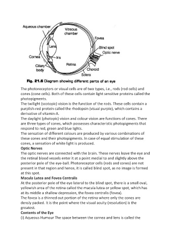
Optic Nerves
The optic nerves are connected with the brain. These nerves leave the eye and
the retinal blood vessels enter it at a point medial to and slightly above the
posterior pole of the eye-ball. Photoreceptor cells (rods and cones) are not
present in that region and hence, it is called blind spot, as no image is formed
at this spot.
Macula Lutea and Fovea Centralis
At the posterior pole of the eye lateral to the blind spot, there is a small oval,
yellowish area of the retina called the macula lutea or yellow spot, which has
at its middle a shallow depression, the fovea centralis (fovea).
The fovea is a thinned out portion of the retina where only the cones are
densly packed. It is the point where the visual acuity (resolution) is the
greatest.
Contents of the Eye
(i) Aqueous Humour The space between the cornea and lens is called the