
Page 2 - LN-CH-16
P. 2
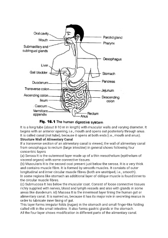
It is a long tube (about 8-10 m in length) with muscular walls and varying diameter. It
               begins with an anterior opening, i.e., mouth and opens out posteriorly through anus.
               It is called canal (not tube), because it opens at both ends [i.e., mouth and anus).
               Structure Wall of Alimentary Canal
               If a transverse section of an alimentary canal is viewed, the wall of alimentary canal
               from oesophagus to rectum (large intestine) in general shows following four
               concentric layers
               (a) Serosa It is the outermost layer made up of a thin mesothelium (epithelium of
               visceral organs) with some connective tissues.
               (b) Muscularis It is the second coat present just below the serosa. It is a very thick
               and contains muscle fibre. It is formed by smooth muscles. It consists of outer
               longitudinal and inner circular muscle fibres (both are unstriped, i.e., smooth).
               In some regions like stomach an additional layer of oblique muscle is found inner to
               the circular muscle fibres.
               (c) Submucosa It lies below the muscular coat. Consist of loose connective tissues
               richly supplied with nerves, blood and lymph vessels and also with glands in some
               areas like duodenum. id) Mucosa It is the innermost layer lining the human gut or
               alimentary canal. It is named so, because it has its major role in secreting mucus in
               order to lubricate inner lining of gut.
               This layer forms irregular folds (rugae) in the stomach and small finger-like folding
               called villi in the small intestine. It also forms gastric glands in the stomach.
               All the four layer shows modification in different parts of the alimentary canal.