
Page 13 - LN-ANATOMY OF FLOWERING PLANT
P. 13
The tissue organisation of roots, stems and leaves can be studied better and
conveniently by the transverse sections of the mature zones of these organs.
I.Dicotyledonous Root
The primary internal structure of dicot root can be studied from the Transverse
Section (TS) of a young root of sunflower, pea or gram. The primary root is the one
which has only primary permanent tissues that are formed from vegetative shoot
apex. Secondary tissues are absent.
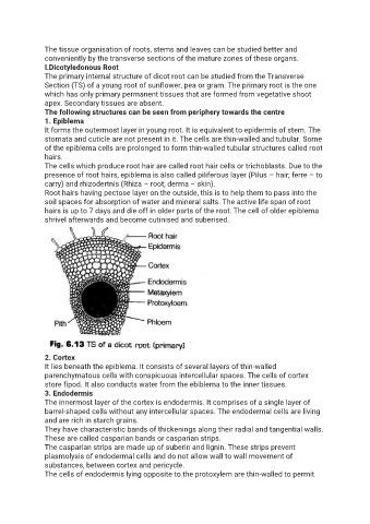
The following structures can be seen from periphery towards the centre
1. Epiblema
It forms the outermost layer in young root. It is equivalent to epidermis of stem. The
stomata and cuticle are not present in it. The cells are thin-walled and tubular. Some
of the epiblema cells are prolonged to form thin-walled tubular structures called root
hairs.
The cells which produce root hair are called root hair cells or trichoblasts. Due to the
presence of root hairs, epiblema is also called piliferous layer (Pilus – hair; ferre – to
carry) and rhizodertnis (Rhiza – root; derma – skin).
Root hairs having pectose layer on the outside, this is to help them to pass into the
soil spaces for absorption of water and mineral salts. The active life span of root
hairs is up to 7 days and die off in older parts of the root. The cell of older epiblema
shrivel afterwards and become cutinised and suberised.
2. Cortex
It lies beneath the epiblema. It consists of several layers of thin-walled
parenchymatous cells with conspicuous intercellular spaces. The cells of cortex
store fipod. It also conducts water from the ebiblema to the inner tissues.
3. Endodermis
The innermost layer of the cortex is endodermis. It comprises of a single layer of
barrel-shaped cells without any intercellular spaces. The endodermal cells are living
and are rich in starch grains.
They have characteristic bands of thickenings along their radial and tangential walls.
These are called casparian bands or casparian strips.
The casparian strips are made up of suberin and lignin. These strips prevent
plasmolysis of endodermal cells and do not allow wall to wall movement of
substances, between cortex and pericycle.
The cells of endodermis lying opposite to the protoxylem are thin-walled to permit