
Page 19 - LN-ANATOMY OF FLOWERING PLANT
P. 19
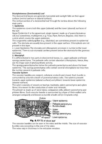
Dicotyledonous (Dorsiventral) Leaf
               The dorsiventral leaves are generally horizontal and sunlight falls on their upper
               surface (ventral surface or adaxial surface).
               The vertical section of a dorsiventral leaf through the lamina shows the following
               main parts
               1. Epidermis
               The epidermis covers both the upper (adaxial) and the lower (abaxial) surfaces of
               the leaf.
               Upper Epidermis It is the uppermost, single layered, made up of parenchymatous
               cell, but sometimes, multilayered, e.g, Ficus, Piper, Nerium, Begonia. Also there is
               cuticle which covers the upper epidermis.
               The outgrowths called papillae {e.g., Gladiolus) are sometimes present in epidermal
               cells. The stomata are usually less present in the upper surface. Chloroplasts are not
               present in this layer.
               ii. Lower Epidermis The stomata and chloroplasts are more in number in the lower
               epidermis. There is sub-stomatal cavities present below the stomata for the gaseous
               exchange.
               2. Mesophyll
               It is differentiated in two parts in dorsiventral leaves, i.e., upper palisade and lower
               spongy parenchyma. The palisade cells contain abundant chloroplasts, Hence, they
               are the major seat of photosynthetic activity.
               The spongy parenchyma lies below the palisade parenchyma and above the lower
               epidermis. This spongy parenchyma cells contain several chloroplasts but less than
               the number present in palisade cells.
               Vascular System
               The vascular bundles are conjoint, collateral, endarch and closed. Each bundle is
               surrounded by a bundle sheath of parenchymatous cells. The xylem is present
               towards upper epidermis (adaxial surface) and phloem towards lower epidermis
               (abaxial surface).
               The xylem consists of vessels or trachae, tracheids, xylem parenchyma and xylem
               fibres. It is meant for the conduction of water and minerals.
               The phloem is made up of sieve tubes, companion cells, phloem parenchyma and
               phloem fibres. Each vascular bundle is surrounded by a layer of thick-walled cells
               arranged compactly and known as bundle sheath cell (in C4-plants only).


               The vascular bundles can be seen in the veins and the midrib. The size of vascular
               bundles vary according to the size of the veins.
               The veins vary in thickness in the reticulate venation.